Subungual Melanoma
What is subungual melanoma?
Subungual melanoma is also termed as nail bed melanoma in which nail morphology becomes defective. The frequency of clinical incidence is not common. The reported incidence revealed that this ailment is not frequent in white skinned people but more predominant in dark skin individuals.
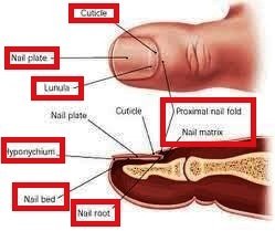
Image 1: Anatomy of Nail
Initially the posterior nail fold become dark and in advanced stage lesion is progressed. Unawareness about the syndrome and ignorance are the common reason behind that the delayed diagnosis in initial stage. People usually consult with doctor, when the lesion formed andrate of healing is negligible.

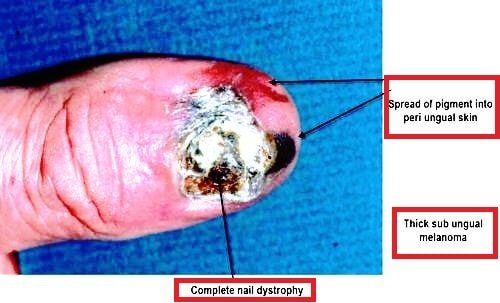
Image 2: Description of Subungual Melanoma
Bacterial or fungal infection is associated with subungual melanoma and often leads to blood clot formation inside the tissue. Mostly thumb or big toe is mostly affected by subungual melanoma, but it can develop in any nail. The incidence of this cancer mostly occurs after 60 years of age.
Subungual Melanoma Symptoms
It is important to remove nail polish and careful examination of the nail bed is important for observation of subungual melanoma, as an initial symptom which includes the dark strip formation on nail bed is covered under the nail police and does not notice. The symptoms include
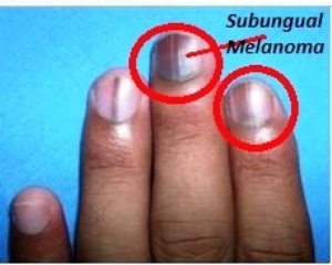
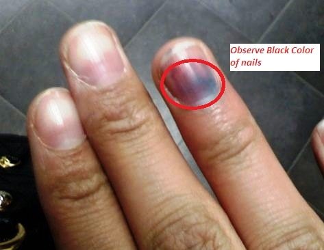
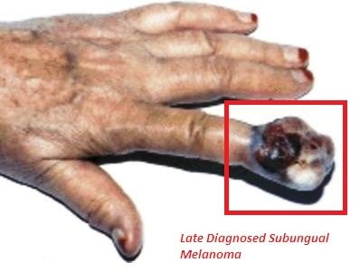
Image 3: Subungal Melanoma
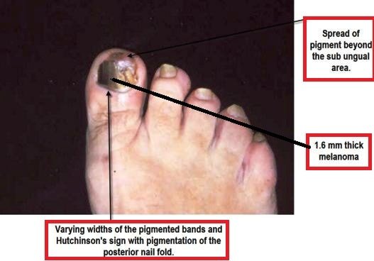
- Approximately greater than 3 mm wide dark band formation in the affected nail.
- The usual colours of the band are black, brown or purple
- Band is rapidly spreading and microscopic examination detects irregular patch formation with colour variation.
- Development of these bands arranged in parallel with each others in a longitudinal direction in the nail bed.
- Gradually papules are formed at the edge of the nail border and turned to plaques. In advanced stage, ulceration is developed. Delayed wound healing causes deformed shape and structure of the nail.
- Sometimes a lump formed under the nail plate with no colour changed
- Affected nail split vertically.
- Nail cuticle becomes affected and white- gray regression occurs.
- Extension of Hutchinson’s sign occurs in advanced stage.
- In some cases, the nail become protruded out
- Sensation of cuticle is become lessened. There may be no pain sensation when the cells present in the nail bed become suppurate or bleed.
Subungual Melanoma Causes
The surveys revealed that physiological and ethnic factors are important for the development of the sublingual melanoma. The incidence rate is high in Asian, African and Native American population.
Direct physical injury and within a specific time period healing has not occurred can cause further infection in the nail bed. Prolonged untreated condition increases the risk of progression of sublingual melanoma. Dermatitides and systemic diseases such as Addison’s and Peutz-Jegher’s are also causes sublingual melanoma.
Other than this, there are three different types of melanoma which can be a cause of development of sublingual melanoma. Melanoma is a type of cancer which is developed in the skin and cancerous growth occurs in melanocytes (pigment cells present in the skin). They are:
- Acral lentiginous melanoma is developed in palms and feet.
- Nodular melanoma, which is formed nodules in the skin and rapidly extended. The growth of the nodules is thickened with time and severity is increased.
- Desmoplastic melanoma is not reported frequently, but it develops due to over pigmentation of the skin from prolong exposure in the UV rays. The diagnosis of this melanoma is difficult as it is similar like other skin lesion.
Subungual Melanoma Treatment
The only option to treat subungual melanoma is surgery. The surgical procedure is conducted for entire tumour expurgation and complexity is present in the surgery. The grade of surgical removal is totally depends upon stage of the melanoma.
In initial stage, when the lesion width is less than 1mm, local expurgation helps to control the growth of the melanoma. In advance stage, expurgation with lymph node biopsy is required to conduction as spreading of the cancerous growth needs to estimate for analyzing the further progression of proliferation. In extremely advance stage of subungual melanoma, some surgeons prefer to complete amputation of the affected finger or toe, but it depends upon extension of the lesion.
Conventional amputation often gives safe and effective results without hampering enduring functional activity. In modern medical technologies has some doubt about complete amputation. If complete removal of cancerous cells is not possible, then chemotherapy needs to implement for complete destruction of cancerous growth.
Prognosis
The different studies help to estimate that the prognosis of subungual melanoma is poor due to late diagnosis. Usually diagnosis is conducted at the late stage of subungual melanoma (SM), when the lesion has expanded more than 4.8 mm area. There is a direct link between tumour expansion and survival rate. In SM, 88% of patients can survive maximum 5 years with 4.8 mm thicken melanoma tumour and only 40 % cases are diagnosed with less than 2.5 mm thickness of the tumour.
Subungual Melanoma Pictures
References
- http://www.dermpathmd.com/cases/melanocytic_proliferations/Subungual%20Melanoma.pdf
- http://www.med.nyu.edu/surgery/oncology/patient-care/melanoma/special-situations/subungual-nail-bed
- http://www.dermnetnz.org/hair-nails-sweat/melanoma-nailunit.html
- http://www.ncbi.nlm.nih.gov/pmc/articles/PMC1243122/
- http://www.cfp.ca/content/54/12/1698.full