Cervical Stenosis
Introduction
Cervical stenosis is a condition wherein there is a narrowing in the spinal column due to several causes. This condition places the patient at risk for an acute neurologic injury because of the greater possibility of spinal cord compression [1].
What is Cervical Stenosis?
The spinal column is a bony tube that is composed of 33 vertebrae and runs from the cranium to the coccyx. It provides support to the back area and houses the spinal cord.
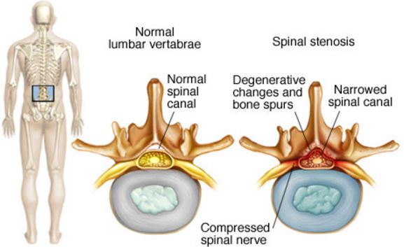
Figure 1 – shows the inside of the spinal column
Cervical stenosis refers to the narrowing of the space inside the upper part of the spinal column [2,3,4].
The narrowing may occur in the center spinal canal, the openings in between consecutive vertebrae or the canals where the nerve exits the spinal column. This condition is considered to be part of the aging process but it may also occur younger people who are born with a narrowing of the spinal canal and those who suffer from a spinal injury [2].
ICD-10 Code of Cervical Stenosis
The 2016 ICD-10-CM diagnosis code used for cervical stenosis is M48.02. This code is for the spinal stenosis of the cervical area or region [7].
Causes of Cervical Stenosis
Primary stenosis
Primary stenosis may occur in individuals who have the following congenital malformation [1, 5].
- Spinal Dysraphism- malformations of the spinal cord
- Segmentation failure
- Achondroplasia- gene mutation that causes abnormal growth of the spine
- Osteopetrosis- osteoclasts are unable to resorb bone
Acquired stenosis
Degenerative changes
Degeneration of the body is viewed to be the most common cause of cervical stenosis. Stress and strain will cause wear and tear to the degenerating spine and this will cause problems.
There are bone spurs that may develop and this will protrude to the spinal canal. The protrusion will decrease the available space to the spinal cord. The collapse of intervertebral discs may also lead to the reduction of the spinal canal space [1, 3].
Tumor growth
Tumors can form in several places in the spinal column. It can either be in the membrane that covers the spinal cord, inside the spinal tube or the space in between the vertebrae and spinal cord. The tumor occupies the space for the spinal cord and may even press on it [6].
Iatrogenic changes
A patient who just underwent surgical procedures in the spine such as discectomy or laminectomy may suffer cervical stenosis due to the swelling of the tissue. The inflamed tissue may put pressure on the nerves or spinal cord [1, 6].
Trauma
Injuries that result from traumatic incidents such as car accidents may cause fractures or dislocations in the vertebrae of the spinal column. A bone may be displaced and compress or damage the spinal cord inside the column [6].
Symptoms of Cervical Stenosis
Many individuals who are more than 50 years old may have some degree of spinal canal narrowing but do not present with any signs or symptoms. The symptoms associated with cervical stenosis only appears once either the nerves or spinal cord is compressed. It is important to note that these symptoms develop slowly over a long period of time [8, 9].
An individual with cervical stenosis may experience:
- Bowel or bladder incontinence (loss of control)
- Neck pain
- Numbness or weakness of the shoulders, arms, hands or legs
- Problems in balance and coordination. Cervical stenosis may lead to paralysis if the spinal cord is damaged.
Diagnosis of Cervical Stenosis
Health history and physical examination
Establishing a diagnosis of cervical stenosis will start in a health history and physical examination. The physician will obtain more information about the discomfort and how it has affected activities of daily living. Any other pain or changes in elimination pattern will have to be looked as well [3, 4, 8,]
A physical examination will be performed next. This exam will identify the neck movements that causes any pain and discomfort along with the skin sensation, reflexes and muscle strength. The physician will also check any problem in walking or balance [2, 3].
Laboratory Tests
An x-ray will be requested in order to visualize the cause of cervical stenosis. It will be able to show any sign of degeneration, vertebral collapse or growth of bone spur that may have caused the narrowing of the spinal canal [3, 8, 9].
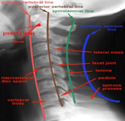
Figure 2 – Normal x ray of Cervical spine
The physician may also request a magnetic resonance imaging (MRI) in order to get a clearer picture of the cause of the problem. An MRI will be able to pinpoint the exact location where the spinal cord is being pressed on [3, 6, 8].
Electrical tests of the nerves of the arm and hand, such as an electromyography, may also be asked to see any possible problem in the motor pathway due to cervical stenosis [3].
Treatment of Cervical Stenosis
Nonsurgical method
If the cervical stenosis of the individual is mild, the symptoms that are experienced may be managed by pain medications and physical therapy.
A Nonsteroidal anti-inflammatory drug (NSAID) is usually given to patients to assist in relieving the pain and inflammation. Muscle relaxants may also be prescribed to alleviate the spasm that may occur in this condition [2, 6, 8].
Physical therapy may be advised to address the other effects of cervical stenosis. Regular sessions with the therapists will prevent muscle weakness and build up the strength and endurance of the muscles. It will also help improve the balance and maintain the stability and flexibility of the spine [6, 8].
Decompressive Surgery
If the condition of the patient continues to progress, a surgical procedure may be advised by a physician. A decompressive surgery is done in order to help make room in the spinal canal or relieve the pressure on the spinal cord.
The procedure is done either from the front or the back of the neck and bone or tissue that presses on the spinal cord is removed. The vertebrae may also be surgically fused together to provide additional stability to the spinal column [8, 9].
References
- Hsiang, J. K. (2015, July 9). Spinal Stenosis. Retrieved from emedicine: http://emedicine.medscape.com/article/1913265-overview
- National Institute of Arthritis and Musculoskeletal and Skin Diseases. (2013, January). Spinal Stenosis. Retrieved from National Institute of Arthritis and Musculoskeletal and Skin Diseases: http://www.niams.nih.gov/Health_Info/Spinal_Stenosis/
- Houston Methodist. (2003). A Patient’s Guide to Cervical Spinal Stenosis. Retrieved from Houston Methodist: http://www.eorthopod.com/Booklet?ClinicID=6138752e01d21f68baaa5b7b82751802&TopicID=f2e57ac35842f72df6f9f61469345c27
- Rhyne, A., & Claytor, B. (2015, June 6). Understanding Cervical Stenosis. Retrieved from Understandspinesurgery: http://www.understandspinesurgery.com/articles/read/understanding-cervical-stenosis
- Johns Hopkins Medicine. (n.d.). Neurology and Neurosurgery. Retrieved from Johns Hopkins Medicine: http://www.hopkinsmedicine.org/neurology_neurosurgery/centers_clinics/pediatric_neurosurgery/conditions/achondroplasia.html
- Mayo Clinic. (2015, November 15). Spinal Stenosis. Retrieved from Mayo Clinic: http://www.mayoclinic.org/diseases-conditions/spinal-stenosis/basics/causes/con-20036105
- (n.d.). Spinal Stenosis, Cervical Region. Retrieved from ICD10data: http://www.icd10data.com/ICD10CM/Codes/M00-M99/M45-M49/M48-/M48.02
- (2014, June 4). Cervical Spinal Stenosis. Retrieved from WebMD: http://www.webmd.com/back-pain/tc/cervical-spinal-stenosis-credits
- University of Maryland Medical Center. (2003). Cervical Spinal Stenosis. Retrieved from University of Maryland Medical Center: http://umm.edu/programs/spine/health/guides/cervical-spinal-stenosis