Ankle Anatomy
About Ankle
The ankle is the joint that is located between the leg and the foot. This joint is a main contributor of stability in the lower limbs and it allows humans to perform actions such as running, jumping and walking [1, 2].
Clinical Anatomy
The ankle joint, also known as talocrural joint, is an example of a synovial joint and is formed by the bones, tendons and ligaments found in the leg and the foot [1, 2].
Bones
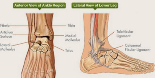
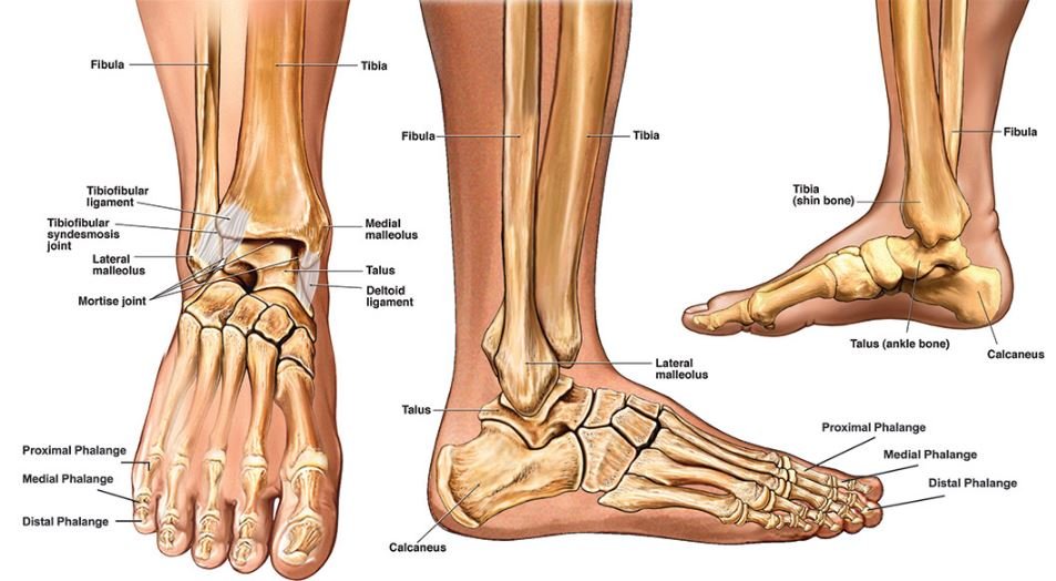
There are several bones that make up the ankle: the tibia, the fibula, the talus and the calcaneus. When viewed anteriorly, the tibia makes up the medial part of the ankle while the fibula forms the medial portion of the ankle. The talus bone and the calcaneus comprise the bottom part of the joint.
The tibia, fibula and the talus bones form what is called the true ankle joint which is responsible for the up and down movement of the ankle. The talus and the calcaneus form the subtalar joint which permits the foot to move from side-to-side. Figure 1 shows the bones of the ankle joint [3, 4].
The ends of the tibia and fibula forms the protrusions in the ankle and have their respective names. The end of the tibia that is on the inside of the ankle is called the medial malleolus and the one protruding at the back is pertained to as the posterior malleolus. The low end of the fibula felt on the lateral side of the ankle is the lateral malleolus [4].
Tendons
Tendons or elastic tissues connect the bones and joints to the muscle. There are several tendons found in the ankle and the largest among these is the Achilles tendon. This tendon attaches from the calf muscle to the heel and allows the body to run, jump and walk up the stairs. Figure 2 shows the Achilles tendon [4, 5].
Ligaments
The ligaments are bands of flexible, fibrous connective tissues that ensure that the tendons are held in place and the joints are stabilized. There are 2 sets of ligaments that originates from the malleolus.
The medial ligament, also known as deltoid ligament, consists of 4 ligaments (tibionavicular ligament, posterior tibiotalar ligament, tibiocalcaneal ligament and anterior tibiotalar ligament) and attaches from the medial malleolus. The said ligaments fan out and connect to the calcaneus, talus and the navicular bones. The medial ligament prevent over-eversion of the foot [5, 6].
The lateral group of ankle ligaments composed of 3 ligaments: calcaneofibular ligament, anterior talofibular ligament and posterior talofibular ligament. They attach to the lateral malleolus and they are smaller than the medial ligament which makes sprains of the lateral ligament to be more common. These ligaments prevent excessive inversion of the foot [6, 7].
Movement
The ankle joint is an example of a hinge joint and joints in this category are only able to move along one axis. The ankle is only able to dorsiflex and plantarflex. Dorsiflexion of the foot is produced by the muscles located at the back of the leg while plantarflexion is caused by muscles in front of the leg [2, 8].
There is more risk for injury when the ankle is in supine position compared to when it is pronated. This is because the shape of the medial malleolus and the medial ligament makes pronation of the ankle more limited. Studies had shown that around 80% of ankle injuries occur when the foot is in supine position [9].
Common Conditions
Ankle Sprain
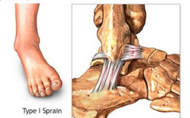
An ankle sprain occurs when the ligaments found in this joint are either partially or completely due to an accidental twist of the foot. This may occur in several sport activities or from a bad landing after a jump [4].
Ankle Fracture
Fractures of the ankle may happen after a fall or a vehicular accident. The severity of an ankle fracture may range from tiny cracks in the bones to breaking of bones into several fragments [4].
Arthritis
Arthritis is the condition where there is inflammation of the joints and the most common types that affect the ankle are osteoarthritis, rheumatoid arthritis and gout. In osteoarthritis of the ankle, the cartilages that cover the ends of the bones break down. Rheumatoid arthritis is an autoimmune condition where the body attacks the joint lining and cause pain and inflammation. Gout is a type of arthritis where uric acid crystals deposit in the ankle joint [4].
Radiology
Radiologic examination of the ankle may be requested by a physician to assist in establishing a diagnosis [4, 9].
X-ray
The x-ray is the most commonly requested radiographic examination because of its availability. This test will be able to show any crack or break in the bones in the ankle [4, 9].
Stress x-ray
In a stress x-ray, the physician will apply pressure on the affected ankle while an x-ray is being taken. This type of x-ray may be able to reveal problems that may not be seen in a regular x-ray [4, 9].
Magnetic Resonance Imaging (MRI)
With the aid of a high powered magnet, the MRI will be able to get images of the ankle. The MRI can help assess the severity of the ankle injury [4, 9].
References
- Healthline Medical Team. (2015, April 8). Ankle. Retrieved from Healthline: http://www.healthline.com/human-body-maps/ankle
- Teach Me Anatomy. (2015, December 15). The Ankle Joint. Retrieved from Teach Me Anatomy: http://teachmeanatomy.info/lower-limb/joints/the-ankle-joint/
- Southern California Orthopedic Institute. (2015). Anatomy of the Ankle. Retrieved from Southern California Orthopedic Institute: http://www.scoi.com/specialties/anatomy-ankle
- Karriem-Norwood, V. (2014, November 15). Image Collection: Human Anatomy. Retrieved from WebMD: http://www.webmd.com/fitness-exercise/picture-of-the-ankle
- Swierzewski, J. (2015, September 15). Muscles, Tendons & Ligaments of the Foot & Ankle. Retrieved from Health Communities: http://www.healthcommunities.com/foot-anatomy/muscles-tendons-ligaments.shtml
- Hoagland, T. (2015, February 25). Ankle Joint Anatomy. Retrieved from eMedicine: http://emedicine.medscape.com/article/1946201-overview#a3
- Sports Injury Clinic. (2014). Ankle Anatomy. Retrieved from Sports Injury Clinic: http://www.sportsinjuryclinic.net/anatomy/ankle-anatomy
- Taylor, T. (2015). Hinge Joint. Retrieved from Inner Body: http://www.innerbody.com/image_skel07/skel31.html
- Goel, A., & Hartley, L. (2015). Ankle radiograph (an approach). Retrieved from Radiopaedia: http://radiopaedia.org/articles/ankle-radiograph-an-approach