Tattoo Infection
What is tattoo?
A tattoo is a permanent design or mark created on the skin by administering ink pigments into the dermis skin layer with the aid of a needle. Tattoo artists utilize a hand portable machine with one or more needles repeatedly driven into the skin layer, acting similarly to a sewing machine.
Ink droplets are imbedded with each needle puncture. When the ink is introduced, the color pigments are absorbed by the underlying dermal cells, which include mas cells, firboblasts and macrophages. The pigmented granules of the tattoo are then located in the intracellular and extracellular layers when viewed through skin light micro-graph usually extending up to 400nm diameter.

Image 1 – Samples of Tattoos on back, right hand.
Tattoo ink bases may be derived from metal salts, iron oxides, plastics, pen ink, blood, and soot. Heavy metals are used to render color which include mercury for red,lead for yellow and green, copper for green and blue, iron for black, red and brown, cobalt for blue, aluminum for violet and green, and barium for white.
Carbon from ash or soot can also be used for black color. The carriers of the solvents are used to aid in the pigment perfusion into the skin layers. Solvents may include water, ethyl alcohol, methanol, glycerin and propylene glycol. Alcohol as a carrier yields better dye transport and helps disinfect the puncture or entry sites.
What is Tattoo Infection ?
The tattooing procedure is commonly performed without the aid of anesthesia. Mild to significant discomfort or pain with minimal bleeding are expected from the process. Majority of tattoos heal without complications. With the invasive nature of the procedure, however, there are risks of infection, contracting bloodborne diseases, allergic reactions, MRI reactions and other skin problems.
The use of tattoo dyes, specifically the colors, yellow, red, blue and green, are associated with allergic response, presented as pruritic lesions even years after the tattoo creation.
Granulomas or skin bumps may develop around the tattoo and soon become keloids. There are also instances that tattoos obscure MRI results, and may even react causing skin burns during radiographic procedures. Unsterile needles can also serve as a vehicle in the transmission of hepatitis B, hepatitis C, and tetanus.
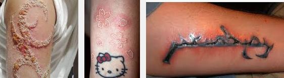
Image 2 – Tattoo Infection of arm, forearm
The breach in skin integrity can lead to possible microbial contamination and subsequent tattoo infection. Higher possibility of infection also occurs with abnormal skin reactions at the tattoo site. The use of asepsis and regular proper tattoo care are paramount to the prevention of infection if properly observed for safety precaution and for post procedural care.
Tattoo Infection Symptoms
Early manifestations of tattoo infection may not be immediately apparent as they mimic the normal healing process. The tattoo area should be observed for the following manifestations:
- Erythema and irritation. Mild redness, discomfort and warmth can be normally felt which should improve in few days. Infection is suspected if the tattoo site becomes more erythematous or reddish with bubbly appearance.
- Fever or pyrexia. Elevated temperature is the hallmark of infection.
- Pain. Mild pain is normal during and after tattoo creation and decreases or resolves in few hours. Worsening pain and shooting pain warrants further evaluation for infection or other problems.
- Inflammation. Although slight swelling is expected, unusual inflammatory response may indicate developing infection.
- Erythematous sores or streaks. The presence of sores or red streaks on the tattoo borders could be reflective of simple infection or worse, staphylococcal infection or even blood poisoning.
- Unusual odor and discharge. The presence of pus, abcess and abnormal odor indicate microbial growth and advancing infection.
Tattoo Infection Causes
Administering foreign dyes or pigments into the skin layer can lead to acquisition of infectious diseases and localization of infection within the tattoo site.
There are tattoo inks and bases which can cause allergic reactions especially to clients with co-morbidities, e.g. immunosuppression, diabetes and cardiovascular disease.
Contaminated needles and inks can cause direct skin infection. To date, the United States Food and Drug administration released a consumer primer last August 7, 2014, warning the public of the potential hazards to health related to a group of tattoo kits which were found to be contaminated with bacteria.
Cultures from infected tattoo lesions usually reveal the presence of any of these microbial agents: Staphylococcus aureus, methicillin-resistant Staphylococcusaureus (MRSA), Streptococcus pyogenes, Clostridium and Mycobacterium haemophilum.
The current hygiene and infection control standards can significantly reduce occurrence of infection, but infection can still be introduced during the breach of the skin layer epidermis. There were also reports of tattoo-related transmission of human immunodeficiency virus (HIV), leprosy, hepatitis and tuberculosis.
Tattoo Infection Treatment
Local bacterial tattoo infection can be treated with topical antimicrobial preparations such as Neosporin, A&D ointment and Bacitracin. Antihistamines and topical corticosteroids are indicated to manage allergic reactions. For severe cases, surgical excision with or without skin grafting is the treatment of choice to remove tattoos.
Traditional laser procedure is not recommended due to the possibility of triggering abnormal systemic reaction. New laser techniques with picosecond laser and/or selective Q-switched lasers are preferred because these allow for the proper matching of appropriate laser wavelengths with the color and base of tattoo pigments used, thus, are safer and lead to lesser scarring.
Example, blue and black pigments can be erased by a laser wavelength of 1064nm, yellow, red and orange with 532 nm wavelength, and green can be removed with a wavelength of 755nm. Whitening or lightening of the skin is immediately noted after laser tattoo removal.
Individuals undergoing tattoo imprinting should also be educated on the proper care of their tattoos to prevent the development of infection. The following precautionary measures should be discussed:
- Bandage is removed after 24 hours.
- Keep tattooed area clean and dry. Avoid swimming until tattooed area is completely healed. Moisture promotes microbial proliferation.
- Religiously apply recommended topical ointment over the site as indicated.
- Avoid tattoo exposure to sunlight during the healing process. Sunscreen should be worn over the tattoo if sun exposure is anticipated.
- Avoid constrictive clothing over the healing site.
References
- Body art.(2015). Infected tattoo Body Art Site. Retrieved from http://www.searchbodyart.com/how-to-treat-an-infected-tattoo/
- Keaney, T.(2015). Tattoo Skin Reactions: Allergies, Infections, and Burns. Medscape Drugs & Diseases from WebMD.. Retrieved from http://reference.medscape.com/features/slideshow/tattoo-skin-reactions
- Linsmeier Kilmer, S., Fitzpatrick, R., and Goldman, M. (2013). Tattoo lasers. Medscape Drugs & Diseases from WebMD.Retrieved from: http://emedicine.medscape.com/article/1121212-overview.
- Mayo clinic. (2015). Tattoos: understand risks and precautions. Mayo Foundation for Medical Education and Research. Retrieved from http://www.mayoclinic.org/healthy-lifestyle/adult-health/in-depth/tattoos-and-piercings/art-20045067
- Rodríguez-Blanco, I,,Fernández, L., Suárez-Peñaranda, J., Pérez del Molino, M., Esteban, J., and Almagro, M. (2011). Mycobacterium chelonae infection associated with tattoos. ActaDermVenereol.91(1):61-2. PMID: 21264454
- USFDA. (2015).Inks Used in Certain Tattoo Kits Cause Infections. US Food and Drug Administration. Silver Spring, MD. Retrieved from http://www.fda.gov/ForConsumers/ConsumerUpdates/ucm316357.htm.
- Wollina, U. and De Cuyper,.C. (2011).Tattoo removal. Evidence Based Dermatology, People’s Medical Publishing House-USA.
- Zachary, C., andRofagha, R. (2012).Laser therapy.Dermatology. 3rd ed. Philadelphia, PA: Elsevier.