Urinalysis
A urinalysis or urine examination is utilized as a diagnostic or screening tool due to its ability to detect the components of the urine that are associated to several pathologic conditions. This can be done as a part of a routine examination or be requested to confirm or establish a diagnosis [1, 2].
What is Urinalysis?
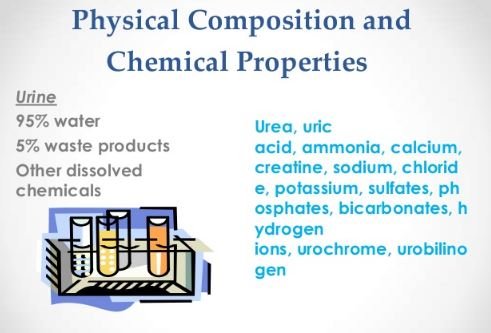
A urinalysis is simply analyzing the urine sample of the patient. In this test, the appearance, concentration and composition of the urine is identified. There is a predetermined set of normal values and any alleviation from this may indicate a pathologic condition. This test only requires around 30- 60 ml of midstream urine and it is either sent to the laboratory for analysis or be tested by a urine test strip. It is essential to have the sample tested right away because the result will be altered if the sample is more than an hour old.
Figure 1 shows the interpretation of the urine test strip [2, 3, 4].
Purpose of Urinalysis
There are several reasons wherein a physician may request for a urinalysis [2, 3, 4]:
Routine Medical Examination
This test is performed as part of the annual physical examination, assessment prior to surgery or hospital admission and screening for any potential kidney problem [3].
Further Examination
When a patient complains of symptoms such as painful urination, abdominal and flank pain, fever or presence of blood in the urine, a urinalysis will be requested to establish a diagnosis of a urinary tract infection [3].
Patient’s Response to Therapy
As the patient receives treatment, the urinalysis will show any improvement or deterioration in his condition. Examples of these pathologic conditions are: uncontrolled diabetes, infection in the kidneys, hypertension and any impairment in the kidneys [3].
Normal Values
Table 1 shows the characteristics of a normal urine [4, 5].
| Parameter | Normal Value |
| Color | Light or pale yellow- dark or deep amber |
| Clarity | Clear or cloudy |
| pH | 4.5- 8.0 |
| Specific gravity | 1.005- 1.025 |
| Glucose | Less than 130 mg/dL |
| Ketones | None |
| Nitrites | Negative |
| Leukocyte esterase | Negative |
| Bilirubin | Negative |
| Urobilirubin | 0.5- 1 mg/dL |
| Blood | Less than 3 RBCs |
| Protein | Less than 150 mg/dL |
| RBCs | Less than 2 RBCs/hpf |
| WBCs | Less than 2 WBCs/hpf |
| Squamous epithelial cells | Less than 15 cells/hpf |
| Casts | 0-5 hyaline casts/lpf |
| Crystals | Occasionally |
| Bacteria | None |
| Yeast | None |
Table 1- Normal Characteristic of Urine
Clinical Significance
Visual Examination
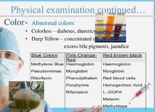
It is the pigment urochrome that determines the color of the urine. Although yellow is the normal color of the urine, it may be lighter or darker depending on the fluid status of the individual. Concentrated urine will be of a darker shade while a diluted urine will have a lighter shade. Other colors such as red, orange or green may be due to a food or drug that is ingested [5].
The clarity of the urine is determined by substances in the urine. The presence of bacteria and high amount of cells and protein may turn the urine turbid [4, 5].
Chemical Analysis
The pH of the urine may be associated with renal stones. Acidic urine is related to uric acid and cysteine calculi. On the other hand, alkaline urine is seen in patients with calcium oxalate and struvite. The specific gravity of the urine is related to the amount of solute and hydration status. A urine with a low specific gravity may be due to diseases such as diabetes insipidus wherein there is a failure to concentrate the urine. The urine’s high specific gravity may be attributed to the high amounts of protein or ketoacids [4, 5].
Increased amount of glucose (Glucosuria) and ketone (Ketonuria) in the urine is seen in uncontrolled diabetes. High levels of glucose overwhelms the kidney and high amounts of it are excreted in the urine. Ketones appear due to fat metabolism secondary to carbohydrate insufficiency [4, 5].
The presence of leukocyte esterase and nitrites in the urine is indicative of a urinary tract infection. Leukocyte esterase is the enzyme produce by WBCs while nitrites are compunds that are produced by gram negative bacteria. Bilirubin and urobilinogen excreted in the urine are possible signs of a hepatic pathology. Proteinuria is possibly associated to the disruption of the barrier that is responsible for the filtration of urine. This symptom is seen in patients with glomerulonephritis and nephrotic syndrome [4, 5, 6, 7].
Microscopic Examination
Increased amount of RBCs in the urine is called hematuria and associated to damage to the glomerulus or kidney trauma. Large number of WBCs, bacteria and yeast may be found in the urine during an infection. Significant amount of epithelial cells may indicate contamination of the urine sample while the presence of casts is an evidence of glomerular inflammation. Crystals can be seen even in normal patients but there are particular ones such as tyrosine or leucine crystals that are only present in those with liver impairment [4, 5].
References
- Labtests Online. (2015). Retrieved from Labtests Online: https://labtestsonline.org/understanding/analytes/urinalysis/ui-exams/start/1#nitrite
- Mayo Clinic. (2014, February 15). Urinalysis. Retrieved from Mayo Clinic: http://www.mayoclinic.org/tests-procedures/urinalysis/basics/definition/prc-20020390
- Nabili, S. (2015, April 30). Urinalysis. Retrieved from Medicinenet: http://www.medicinenet.com/urinalysis/article.htm#what_is_a_urinalysis
- The Internet Pathology Laboratory. (2002). Urinalysis. Retrieved from The Internet Pathology Laboratory: http://www.health.auckland.ac.nz/webpath/tutorial/urine/urine.htm
- Lerma, E. (2015, December 16). Urinalysis. Retrieved from Emedicine: http://emedicine.medscape.com/article/2074001-overview#a2
- Health Guide. (2013, August 18). Leukocyte Esterase Urine Test. Retrieved from New York Times: http://www.nytimes.com/health/guides/test/leukocyte-esterase/overview.html
- (2015, October 21). Nitrates or Nitrites in Urine. Retrieved from Minimedia: http://minimemedia.com/nitrates-in-urine/