Supracondylar Fracture
What is supracondylar fracture?
Supracondylar fracture means elbow fracture. It is more common in young age children rather than adult individuals. Humerus, radius, and ulna are the three upper limb bones which joint together at Supracondylar (elbow) joint. Associated ligaments, muscles, nerves and blood vessels are often also injured in supracondylar fracture.
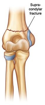
Image 1: Supracondylar fracture
The reported case studies revealed that 10% of total fracture occurs in children is supracondylar fracture. The incidence is more frequent in boys at the age of 5 to 7 years. During this age the structures associated with supracondylar joint are continuing for transformation. Usually for this reason the plate of the joint is thinner and more susceptible to fracture during any accidental trauma.
Supracondylar Fracture Types
There are different types of supracondylar fracture and their classifications are as follows:
Fracture occurs in the upper region of supracondylar (elbow). This is the commonest supracondylar fracture occurs in the lower part of the humerus (arm bone) where they attached with an elbow. Children below the age of 8 years, mainly get this type of fracture, as their humerus is not strong enough. This may become a serious injury if the underlying nerves become injured.
Fracture at the protuberance of elbow. Bony protuberances are present at the supracondylar (elbow) joint. Any of them may get fractured during injury, but outer and lateral tip most frequently fractured. Usually a growth plate and joint surface both has been broken.
Fracture at the tip of the elbow. The internal medial tip of each protuberance may be fractured during the age of 9 to 14 years of a child during accidental fall down. Tip of each protuberance is termed as epicondyle.
Fracture in Growth plate. Growth plates are situated at the end of the humerus, radius and ulna bones where they connected with ligaments. Growth plate establishes hand structure for the adult bone. Fracture at the growth plate may hamper the structure of the hand.
Fracture in radius or ulna. The usual affected bone is radius, in excess of load breaks the head of the radius and even compression fracture also occurs at radius. Generally tip of the ulna may affect, but reported case is unusual.
Fracture in ulna with associated dislocation. Ulna fracture can also dislocate the upper part of radius which is attached to elbow joint. Fracture repair with careful treatment for dislocation is required to achieve complete restoration.
Open fracture. In open fracture skin is also tearing up and associated muscles, tendons, and ligaments also become injured. Recovery is usually taken a longer time.
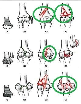
Image 2: Types of Supracondylar fracture
Symptoms
Common symptoms arise with any types of fracture in upper limb gives following symptoms
- Unbearable pain
- Inflammation of the elbow joint
- Lack of sensation in the hand arises with nerve damage
- Arm movement is restricted.
Causes
Children usually involved in different types of physical activities and they are careless about the danger associated with jumping, running or other sports activities.
The fracture in elbow joint occurs due to following reason
- Outer stretching of the arm during fall down or other physical activities
- Fall down and extreme load occurs at the elbow
- Direct injury at the elbow.
Diagnosis
Diagnosis is usually based on physical examination and for confirmation X-ray is conducted. It is important to diagnose the nerve or blood vessels injury are take place or not with fracture. The following sign helps to identify the fracture.
- Inflammation
- Injury at the site of elbow
- Unable to hand movement
- Warmth
- Blood clotted due to hampered circulation.
X-ray helps to estimates the structures which are affected due to injury. An anteroposterior and a lateral view of elbow joint help to diagnose the exact location of fracture and associated dislocation. Sometimes both hands X-ray conduction is important to evaluate the shape of the arm bones..
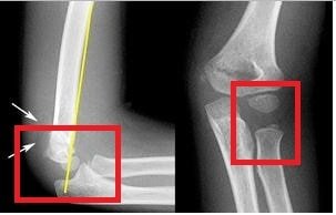
Image 3: X-Ray Image of Supracondylar Fracture
Supracondylar Fracture Treatment
- The initial step of treatment is stabilization of limb and for that purpose application of splint is useful.
- Well padded splint is recommended for multiple fracture or open fracture.
- Cast is applied for restrict the mobilization and helps to hold the bones in a proper position without displacement during healing period.
- Fracture with displacement needs to repositioning of bones. The process of relocating the bone or bones in their original place is termed as closed –reduction. Addition of sedatives or anaesthetics assists to lessen the pain during the procedure.
- Closed surgical reduction and pinning is also recommended if the bone or bones are fragmented and displaced from their original position.
- Open surgery and internal fixation is also required for open fractured associated with nerve or blood vessels damage.
Complications
The following complications may arise during or later stage of fracture:
- Traumatic and iatrogenic nerve injures specially the anterior interosseous nerve and radial nerves are affected.
- Vascular damage causes inadequate blood supply to the arm.
- In distal part of the upper arm bone may develop angular malunions.
- The most common difficulty arises in the later stage of supracondylar fracture is Cubitus varus, or ‘gun-stock deformity’.
References
- http://radiopaedia.org/articles/supracondylar-fracture
- http://emedicine.medscape.com/article/1269576-overview
- http://orthoinfo.aaos.org/topic.cfm?topic=A00037
- http://www.ncbi.nlm.nih.gov/pmc/articles/PMC2682409/