Sialolithiasis
What is Sialolithiasis?
Sialolithiasis is a disease associated with salivary gland. Usually crystalline mass which looks similar like calculus is formed mostly (92%) in the submandibular gland. The affects is mostly developed inside the duct rather than parenchyma. The other salivary glands, which include parotid gland and sublingual gland are rarely affected.
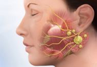
Image 1: Anatomical description of salivary glands
The incidence rate is high in adult male at the age between 30 to 60 years. Children are less commonly affected. Acute and chronic infection can be a cause of development of the sialolithiasis. The development of the calculus in the salivary gland obstructs the saliva secretion. This is not a serious health issue, but develops an oral discomfort. Sialolithiasis size and shape vary and it may be single or multiple. Generally submandibular sialolithiasis is multiple and larger in size. The usual composition of calculi is calcium phosphates and organic matrix.
Symptoms
The main function of the saliva is moist the oral cavity and it also helps to swallow of the food.
- The major symptom after the occurrence of sialolithiasis is a pain in the oral cavity which worsens during and after taking meal.
- The pain can extend up to face and even at the neck
- Difficulty is swallowing.
- Inflammation and tenderness occur in the oral cavity.
- Dryness inside the mouth
- Sluggish saliva movement causes infection at the site of calculus formation
- Redness in the affected area
- Bad smell inside the mouth
- Fever
Causes
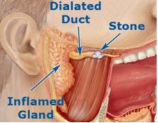
Image 2: Sialolithiasis
The cause of development of sialolithiasis is divided into two aetiologies:
- Saliva preserved inside the duct due to the flow of the saliva becomes slow and in opposition to the gravity which increases the strain inside the salivary duct.
- Composition of saliva. It is considered that when saliva gets supersaturated or deficiency of crystallization inhibitor. The flow of the saliva is hindered due to super-saturation of the saliva. Deficiency of crystallization inhibitor promotes the calcium phosphates and organic matrix such as high mucin composite and calculi formed. The saliva becomes alkaline in nature.
- Malnutrition or an eating disorder in which person not taking sufficient food causes reduction of saliva secretion which leads to sialolithiasis.
- Dehydration or less fluid intake concentrates saliva composition and sialolithiasis occur.
- Some medications such as antihypertensive drugs, anti-allergic and anti-cholinergic drugs reduce the saliva production in the gland and sialolithiasis may occur as the side effect of the drug.
Diagnosis
Diagnosis of sialolithiasis needs to conduct a physical examination, blood test and ultrasound of the oral cavity, Facial X-ray and CT scan.
Physical examination
Usual findings on physical examination are:
- Facial swelling which may extend from neck to eye.
- Peri-orbital oedema
- Tenderness in the neck
- Palpation at the affected side of the neck
Blood Test
Usually following blood test is conducted to estimates the infection and the erythrocytes volume.
- Total WBC count (TLC)
- Red Blood Cell Count
- Haemoglobin estimation
- Hematocrit blood testing
- Mean corpuscular volume (MCV)
Imaging Techniques
Ultrasound of oral cavity helps to detect the sialolithiasis and multiple radiographic images obtained from the CT scan and X-ray help to diagnose the extension, size, shape, location and number of calculi.
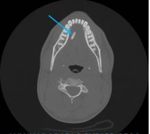
Image 3: Radiographical overveiw of sialolithiasis
Treatment
Treatment of sialolithiasis depends upon the size and location of the calculi.
- Some natural remedies help to treat sialolithiasis such as application of hot compression, mild massage on the affected part, more fluid intake and citric acid drops or lemon drops. All these approaches apply to move the calculi and increase the saliva production, which forcefully rid off the calculi. Lemon drops help to break down the calculi.
- To treat the infection, doctors recommend antibiotics.
- For pain reduction analgesics are prescribed.
- Extracorporeal shock wave lithotripsy is conducted by using high-energy sound waves which increase the shock and break the calculi.
- Large size of calculus cannot be broken or removed easily and needs traditional surgical intervention.
References
- http://www.nature.com/bdj/journal/v193/n2/full/4801491a.html
- Salivary Duct Stones: Symptoms, Causes & Diagnosis at http://www.healthline.com/health/salivary-duct-stones
- https://vula.uct.ac.za/access/content/group/ba5fb1bd-be95-48e5-81be-586fbaeba29d/Sialolithiasis%20and%20sialendoscopy.pdf
- Sialolithiasis: Radiological Diagnosis and Treatment at http://eradiology.bidmc.harvard.edu/LearningLab/central/Kakarala.pdf
- Current opinions in sialolithiasis diagnosis and treatment at http://www.ncbi.nlm.nih.gov/pmc/articles/PMC2639864/