Subserosal Fibroid
What is Subserosal Fibroid?
Fibroids are non-cancerous tumors that abnormally grow in a woman’s uterus. There are four different types of fibroids that can develop in different locations in the uterus and these are; subserosal, intramural, submucosal, and pedunculated fibroids.
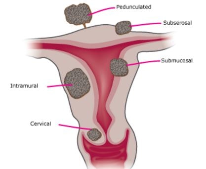
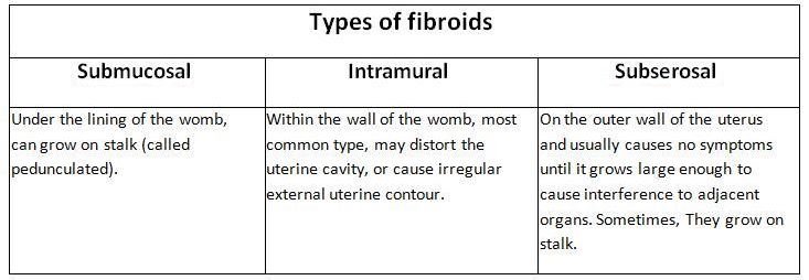
Subserosal fibroids usually develop on the outside of the uterus, which is referred to as serosa. These tumors may grow continuously until it becomes large enough to make the womb look larger on one side. The subserosal fibroid growths will also put extra force on the organs around it.
Sometimes, these tumors might be joined to the uterus by a stem-like or long stalk base. Over time, these fibroid tumors can grow bigger but they do not usually influence the size of the uterus’ cavity. Subserosal fibroids also spread in a specific area among women in their prime reproductive years.
What are the Symptoms of Subserosal Fibroids?
In most women with subserosal fibroids, they do not experience any signs or symptoms. Problems are usually caused by huge and pedunculated fibroid tumors and since these tumors are located outside of the uterus, they do not affect menstrual flows.
Symptoms that are commonly encountered by women with subserosal fibroids include:
- Back pain if it causes a pressure on the spinal nerves
- Pelvic pain once the tumors press on the rectum
- Pain and cramping in the abdomen
- Urinating repeatedly
- Bloating and constipation
- Damaged kidney caused by the ureter compression
- A feeling of heaviness
- Pain may be felt whenever the pedunculated subserosal fibroids twist sometimes
What Causes Subserosal Fibroids?
Physicians still do not know the primary cause of uterine fibroids but according to researchers and other clinical experiences, there are several factors that may have an impact on their formation. These include:
- Hereditary – If a family member has a history of subserosal fibroids, another member of the family may develop it as well.
- Pregnancy – During a women’s pregnancy, the production of progesterone and estrogen normally increases. This might cause the tumors to develop and rapidly grow in size between pregnancies.
- Hormones – Progesterone and estrogen are hormones that trigger the regeneration of the uterine lining during a menstrual cycle and can prompt the growth of subserosal fibroid tumors.
- Other factors – Some substances that help the body preserve tissues like insulin-like growth factor can possibly influence the growth of fibroids.
How is Subserosal Fibroid Diagnosed?
Subserosal fibroids are usually seen by chance during a routine pelvic exam. The physician might feel some irregularities by how the uterus looks like, suggesting the presence of fibroid tumors. If any symptoms had been experienced, the physician may perform some tests and these include:
- Laboratory tests
- Ultrasound – If any confirmations are needed, the physician will do an ultrasound. High-frequency sound waves are used to visualize images of the uterus on a screen. The physician will then see the internal structures and whether fibroids are really present.
- A transducer device is used by the physician which is inserted inside the vagina or just over the abdomen to get images of the uterus.
How is Subserosal Fibroid Treated?
Unluckily, there is no specific medical treatment for subserosal fibroids yet. Medical therapy is only used to ease the symptoms caused by the fibroid tumors and all of the medical treatments are temporary.
Other possible treatments include:
Surgery
This treatment is the most effective for cases of subserosal fibroids. Since the tumors are arranged on the surface of the uterus, laparoscopy is believed to be the most practical way of removing the fibroids. This involves the incision in the abdomen in order to access the uterus and finally removing the fibroids.
Forced ultrasound surgery (FUS)
This is a non-invasive treatment that is proven to be safe and effective. The physician does not need any incisions but with the use of an ultrasound transducer focusing on the sonication to destroy the areas of fibroid tissues.
References
- Uterine Fibroids, Intramural Fibroids, Submucosal Fibroids, Subserosal Fibroids, Breast Fibroids, Fibroid Symptoms, Diagnosing Fibroids, Cancer and Fibroids, Soy And Fibroids, Risk Factors, Fibroid Facts, Dairy Products, Obesity And Fibroids, Uterine Fibroids, Fibrocystic Breasts, Fibroids and Pregnancy, Fibroids And Fertility Fibroid Treatment. at http://www.womens-health.co.uk/suberosal.html
- Uterine Fibroids : Overview,,Types, Causes, Risk Factors, Symptoms, Diagnosis, Treatment, Outlook at http://www.healthline.com/health/uterine-fibroids#Treatment7
- https://ask4ufe.com/what-are-uterine-fibroids/types-of-fibroids/#/subserosal
- Medikare, V; Kandukuri, LR; Ananthapur, V; Deenadayal, M; Nallari, P (July 2011). “The genetic bases of uterine fibroids; a review.” Journal of reproduction & infertility 12 (3): 181–91.
- Okolo S (2008). “Incidence, aetiology and epidemiology of uterine fibroids”. Best practice & research. Clinical obstetrics & gynaecology 22 (4): 571–588.
- Wallach EE, Vlahos NF (August 2004). “Uterine myomas: an overview of development, clinical features, and management”. Obstet Gynecol 104 (2): 393–406.